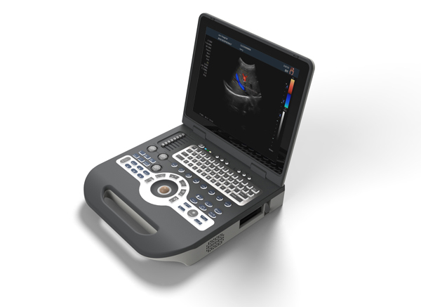
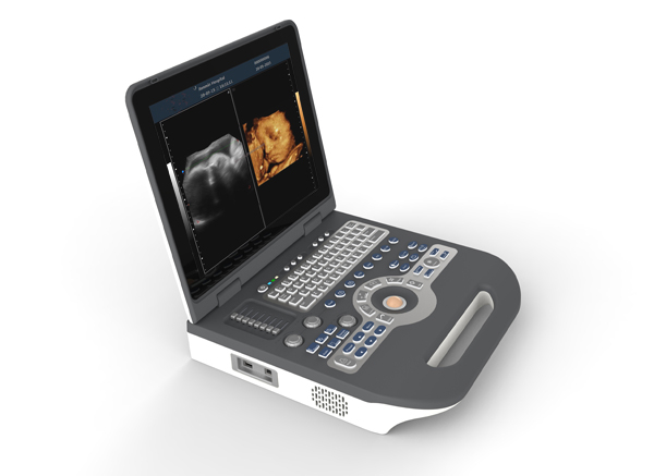
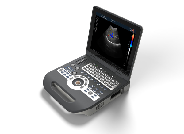
This is a laptop full digital color ultrasound diagnosis system,integrand embedded XP platform performance of stable and affordable.It is a research and development of XF technical team combined with market demand in the XF7800 type main based on the development and design new style,thin and light, good carrying, appearance is compact, the function is powerful, with types of probe can support more, image processing and measuring package software is rich, full blood flow image clarity, auxiliary functions such as adding PW envelope and three imaging modes and practical features such as real-time synchronization.It is widely used in the examination of abdomen, gynecology, obstetrics, urology,vascular, heart, small organs,andrology,musculoskeletal especially in the clinical examination of large medical institutions and primary medical system.Is a cost-effective color doppler ultrasound diagnostic instrument.we have hospital and clinic project solution package, support you complete purchasing job. If any interest, welcome consult details
notebook full digital color ultrasound diagnosis system

Configuration:
1)Full-Digital 2D gray scale imaging
2)Full-Digital Tissue Harmonic Imaging (THI)
3)Color Doppler blood flow imaging
4)Directional color energy Doppler imaging
5)Pulse Wave Doppler imaging (PW)
6)Space compound imaging
7)2D, color, Doppler mode automatic optimization adjustment technology
8)Real-time triple synchronizing
9)Support 6 kinds language
10)Adaptive speckle suppression technology
11)Hand free 3D imaging(Optional)
12)Real-time 4D imaging(Optional)
13)Intelligent picture - in - picture imaging mode (PIP)
14)Monitor: 15 inch high resolution medical LCD monitor, adjustable angle
15)Probe connectors: ≥2 active
Multiple probe configuration:
1.1 Convex probe frequency: 2.0-5.0MHZ(multi-frequency, Harmonic frequency≥5 ), probe scanning angle 20°~85°, visible and adjustable.
1.2 Linear probe frequency: 6.0-12.0MHZ(multi-frequency, harmonic frequency ≥4 ), probe scanningwith trapezoidal imaging technology and 2D beam deflection technology
1.3 Trans-vaginal probe frequency: 5.0-8.0MHZ(multi-frequency, harmonic frequency ≥2 ), probe scanning angle 20°~160°, visible and adjustable.
1.4 Real time 3D (4D) volume probe frequency: 2.0-5.0MHz, 4 segments multi-frequency.
1.5 Micri-convex probe frequency: 4.0-6.0MHz, 3 segments multi-frequency.
Applications: abdominal, urology, OB&GYN, paediatrics / neonatal, superficial / small organ, musculoskeletal, cardiology etc.
Portable color doppler ultrasound diagnostic system

Main technical specification
1. 2D,B/M,PDI,PW,CFM,4D, imaging mode
2. Gray scale: 256
3. Gray Map: ≥16 level, visible and adjustable
4. Dynamic range: 60-240db(visible and adjustable)
5. Resolution: Horizontal≤1mm; Vertical≤0.5mm
6. Under B mode, focus number: 1-6, focus position continuously adjustable
7. STC gain control ≥8 segments
8. THI: harmonic frequency ≥2 segments
9. Line density: ≥256, visible and adjustable
10. Preset: ≥40 kinds, users can customize the inspection conditions for the optimized images of different organs
11. Max scanning depth: ≥775px, visible and adjustable
12. Scanning angle: 50°-100°, visible and adjustable
13. Cine loop ≥4800 frames
14. Adaptive speckle suppressio: 0-100 adjustable
15. Amplification: overall amplification, local amplification, M-type amplification(do M-type sampling amplification under both scanning or freeze state)
16. Color gain: adjustable
17. Color frequency: ≥3 kinds, visible and adjustable
18. Sampling frame: size and position adjustab
19. PW blood flow measurement speed: min speed: ≤0.2 cm/s, max speed: ≥37500px/s
20. PW Doppler frequency: ≥3 kinds, CW Doppler frequency: ≥15 kinds, visible and adjustable
21.Real-time automatic Doppler envelope mapping and automatic measurement and analysis
Measurement and analysis:
1 General measurement
2 OB&GYN measurement
3 Cardiac function measurement and analysis
4 Doppler blood flow measurement and analysis
5Peripheral blood vessel measurement and analysis
6 Urology measurement and analysis
7Orthopedic measurement and analysis
8Automatic Doppler flow measurement and analysis
9Users can programme protocol numbers, formulas and tables
7.1 Diagnostic report editable, embed the ultrasound diagnostic image in the report, and print directly
7.2 Hard disk static and dynamic image storage 120G capacity
7.4 Input / output interface: HDMI port, video input/output port, S-VGA, print port, DICOM 3.0,USB port
notebook full digital color ultrasound diagnosis system

Main Technical Indexes:
The performance requirements of gray-scale imaging mode
The color ultrasonic at the gray-scale imaging performance mode should comply with the provisions of the table 1.1
Table 1.1 At the Gray-scale imaging mode the performance of the probe
performance indexes | probe type and nominal frequency | |||
2.0≤f<4.0 | 2.0≤f<5.0 | 5.0≤f<8.0 | 5.0≤f<12.0 | |
a) probe type and model | Micro-convex | Convex array (type TC60A) | Cavity (type TC10A) | Linear array (type TL40A) |
b) nominal frequency (MHz) | 5 | 3.5 | 6.5 | 7.5 |
c) Scan depth(mm) | ≥90 | ≥160 | ≥40 | ≥50 |
d) Lateral resolution (mm) | ≤3(depth≤50) ≤4(80<depth≤60) | ≤3(depth≤80) ≤4(80<depth≤130) | ≤2(depth≤30) | ≤2(depth≤40) |
e) Axial resolution (mm) | ≤2(depth≤50) | ≤2(depth≤80) ≤3(80<depth≤130) | ≤1(depth≤40) | ≤1(depth≤50) |
f) Blind area (mm) | ≤7 | ≤5 | ≤4 | ≤3 |
g) Transverse geometry precision (%) | ≤20 | ≤15 | ≤10 | ≤10 |
h) Longitudinal geometric location accuracy (%) | ≤10 | ≤10 | ≤5 | ≤5 |
i) Slice thickness (mm) | ≤5 | ≤5 | ≤5 | ≤5 |
j) Perimeter and area measured deviation (%) | ≤±20 | ≤±20 | ≤±20 | ≤±20 |
k) M mode time display error (%) | ≤±10 | ≤±10 | ≤±10 | ≤±10 |
The performance requirements of color Doppler imaging mode:
a). The color Doppler imaging mode should comply with the provisions of thetable 1.2;
b). Color blood flow image should be essentially coincident with the gray-scale image of pipe's;
c). Blood flow direction should be able to correctly identify, no aliasing phenomenon;
Table1.2 at the color blood flow imaging mode the performance of the probe
Doppler model | Micro | Convex array | Cavity | Linear array |
Investigation depth at Color blood flow model | ≥90mm | ≥100mm | ≥40mm | ≥50mm |
Investigation depth at Doppler spectrum model | ≥90mm | ≥100mm | ≥40mm | ≥50mm |
Blood flow speed reading error | ≤±15% | |||

The performance requirements of Doppler spectrum mode:
a). The color ultrasonic at the color Doppler spectrum mode should comply with the provisions of the;
b). Blood flow speed reading error should comply with the provisions of the table2.3;
c). Pulse wave Doppler mode sampling area cursor position should be accurate;
Displayer: 15 inch LCD color display
Running hours: ≥8h;
Input power: ≤300VA;
Host weight: about 6 kg;
Host appearance size: 370 ×382×90(length × width × height) (mm3).
Standard Configuration:
1. One host machine
2. One Li-battery
3. One convex array probe:F=3.5MHZ
One Linear probe: F=7-12Mhz
One trans-vaginal probe: F=5-8Mhz
4. One power adapter
5. Two USB port
6. Svideo output port