I.Specification:
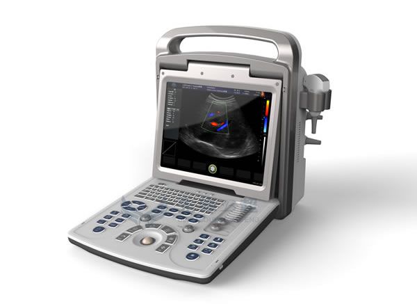
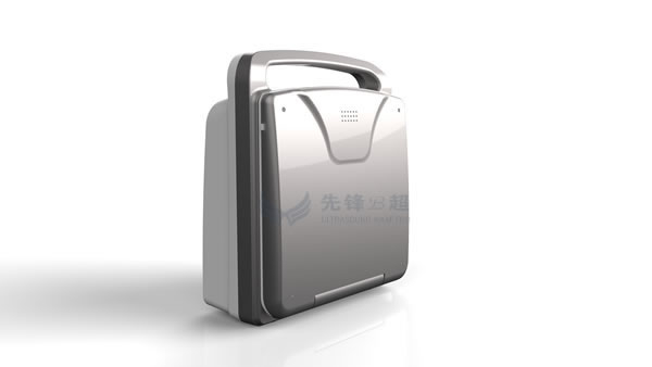
| Model | E20 Portable Color Doppler Ultrasound Scanner |
| Screen Size | 15'LED |
| Application | 1.1 The parameters of probe scanning:electronic convex array; electronic linear array, intracavity electronic micro convex array, electronic micro convex array, electronic phased array probe. 1.2 The mode of operation:B mode,M mode, Color Doppler flow mode(CFM), Power doppler mode(PDI), pulse wave spectral Doppler(PW),tissue harmonic imaging, Real-time two-dimensional/color double mode, Real-time two-dimensional/doppler double synchronous, Real-time two-dimensional/Doppler/ color three synchronization, trapezoid Imaging, Four-dimensional imaging.(4D, optional) 1.3 Interface for probe: four activate probe interface. 2. Ultrasonic imaging technology 2.1 High precision digital continuous beam shaper, fine ultrasound beam control and remove sidelobe noise effectively,improved spatial resolution and contrast resolution drastically, exquisite display full organizational structure. 2.2 Dynamic frequency fusion imaging technology: Automatic adaptive control near field to far field emission, received frequency,making Strong penetration and high-definition image combination perfectly. 2.3 High precision delay receive focus dynamically point by point: it delay the whole image focusing precisely point by point, Present true, delicate organization information. 2.4 Ultra Wide Band imaging technology:you can choose the best center frequency follow your Inclinations according to speciality of different crowds. 2.5 Adaptive technology of image optimization: according to reception of the organization signal at present, automatically digital parameters optimization to render more perfect ultrasonic images. 2.6 Adaptive vessel imaging technology:in allusion to received the doppler echo signal at present, adopt suitable filtering scheme automatically, suppress clutter spectrum effectively, so it achieves the perfect combination of resolution and sensitivity, and effect of blood flow images is much better. 2.7 Adaptive doppler imaging technique: intensify the weak Doppler signal to strong signal,and Strengthen the spectrum signal through processing technology of complex digital. Make the effect of doppler sensitivity and display is improved well. 2.8 THI(Tissue harmonic imaging technology): launch ultrasonic at a relatively low frequency, receives the echo signals of high frequency second harmonic to imaging, at the same time of guarantee good penetration, enhanced tissue imaging resolution and eliminate the fake image maximumly. 2.9 Accurate SRI adaptive suppress speckle and noise: based on the algorithm of accurate image recognition adaptive suppress speckle and noise, not only can smooth spot and noise at the same time also can retain the organizational structure characteristics. 3. measure/ calculate 3.1 General measure B mode: measure, area(method of loci,method of Rectangle , method of ellipsoid ), perimeter(method of loci,method of Rectangle , method of ellipsoid), volume (method of double Plane ,method of area and length, method of ellipsoid), angle, rate, rate of stenosis. M mode: slope, rate, rate of stenosis, heart rate, time. D mode: Doppler measurement for blood flow, Flow velocity, acceleration, pressure gradient, time, velocity integrals, pulsatility index (PI), resistance index. 5.2 Measure and analysis for gynecology: Uterus, endometrium, ovary, cervix uteri, measure of follicle. 5.3 Measure and analysis for obstetrics: GS(Gestation Sac), CRL(Crown Rump Length), LV(Length of the spine), BPD(Bipartitely Diameter), OFD(diameter of occipital and frontal), HC(Head girth), TAD(diameter of transverse abdominal), LVW(width of the lateral ventricle Angle), HW( width of brain hemisphere), TCD(The cerebellum transverse diameter), IOD(diameter of orbital), OOD(distance of Outside the eye),BD(Eyes distance),APTD(Diameter at breast height front and back), TTD(The transverse diameter at breast height), AC(abdominal girth), APD(Abdominal diameter front and back), FTA(Trunk cross-sectional area),HL(length of Humerus ),ULNA(length of ulna),RAD(length of radius),FL(length of femur),TIB(length of tibia ),FIB(length of fibula), APTDxTTD, CLAV(length of clavicle), Hip dysplasia(Hip Angle measurement),etc. it can count gestational age, fetal weight, expected date of confinement,etc. 5.4 Measure and analysis for urology: Prostate volume,bladder volume,residual urine volume,measure and evaluate for angle of coxae (diagnosis of coxae dislocation for newborn ), section Measure (V-Slice). 5.5 Measure and analysis for Small organ and periphery vessel: It mainly used for measure and analysis for cross section area of vessel, heart rate, Stroke Volume, Flow per unit time, ejection time, rate of stenosis, average velocity for blood stream, RI(resistance index), PI( pulsatility index). 5.6 Measurement Report: measurement report for Obstetric, measurement report for gynecology, measurement report for heart, measure for urinary and other,etc, automatic storage measurement results and generate reports. 5.7 Position marker With the probe location more than 95, it can Speedily choose position marking 、Text markup through the intuitive body position detail interface,and it Can preinstall text content; arrow mark,it Supports multiple direction of arrow mark, and is adjustable. |
| Electrical Power | Voltage: 100 V~240 V |
| Power: DC19V 10A | |
| Frequency: 50/60 Hz | |
Physical Specifications | Equipment Size: 390mm(Length)x200mm(Width)x470mm(Height) |
| Equipment weight: 10kg | |
| Conditions | Operating Temperature:5℃~40℃ |
| Humidity:≤80% | |
| Pressure:700hPa~1060hPa | |
| Storage Temperature:-5℃~40℃ | |
| Humidity:≤80% | |
| Pressure:500hPa~1060hPa | |
| Connectivity/Media/Peripherals | Transducer Ports:1 |
| USB Ports: 2 | |
| Hard Disc: 120GB(SSD), 120G/256GB SSD (Optional) | |
| Foot switch: USB | |
| External Display: VGA,HDMI | |
| USB Printer | |
| Digital Laser Printer | |
| Digital B/W Thermal Printer | |
| Cine/Image Memory | Cine Memory: 1024 frame |
| Cine Review Speed: 1,2, 4, 8 | |
| Cine Review Loop | |
| Cine Capture Function | |
| DICOM Connectivity | DICOM3.0 Compliant |
| Image Storage | Storage Format: PNG, AVI, BMP, JPEG, DICOM |
| Export Video Format:AVI | |
| Export Image Format:PNG, JPEG, BMP | |
| DICOM | |
| USB Flash Drive |
II.Function:
Zoom: 10 times
Cine loop: up to 6400 frames
HDD 120GB
III.Standard Configuration:
1.One host machine
2. One convex array probe:F=3.5MHZ
3. USB port
IV.Optional probes: